Cochlea Microscope Slide Labeled . Web the inner ear detects sound, acceleration and position. Be able to recognize and distinguish the auditory parts of the inner ear (cochlea, three different. Back section of slide, behind the cochlea. Web the cochlea is a component of the labyrinth of the internal ear that is responsible for hearing. It consists of fluid filled sacs (membranous labyrinth) that lie in cavities. The cochlea, part of the osseous labyrinth, consists of a long, hollow tube that is coiled into a spiral, resembling a snail. Web be able to recognize them at the light microscope and em level. Web cochlea the cochlea houses an elaborate configuration of membranous labyrinth and hair cells, called the organ of. Web an illustration and a histological section of a single turn of the osseous cochlea show the cochlear duct positioned between the scala vestibuli and scala. Study with quizlet and memorize flashcards containing terms.
from ar.inspiredpencil.com
Back section of slide, behind the cochlea. Web an illustration and a histological section of a single turn of the osseous cochlea show the cochlear duct positioned between the scala vestibuli and scala. Be able to recognize and distinguish the auditory parts of the inner ear (cochlea, three different. Web cochlea the cochlea houses an elaborate configuration of membranous labyrinth and hair cells, called the organ of. It consists of fluid filled sacs (membranous labyrinth) that lie in cavities. Web the inner ear detects sound, acceleration and position. Web be able to recognize them at the light microscope and em level. Study with quizlet and memorize flashcards containing terms. Web the cochlea is a component of the labyrinth of the internal ear that is responsible for hearing. The cochlea, part of the osseous labyrinth, consists of a long, hollow tube that is coiled into a spiral, resembling a snail.
Cochlear Model Labeled
Cochlea Microscope Slide Labeled The cochlea, part of the osseous labyrinth, consists of a long, hollow tube that is coiled into a spiral, resembling a snail. Web the inner ear detects sound, acceleration and position. Web the cochlea is a component of the labyrinth of the internal ear that is responsible for hearing. Back section of slide, behind the cochlea. Web an illustration and a histological section of a single turn of the osseous cochlea show the cochlear duct positioned between the scala vestibuli and scala. The cochlea, part of the osseous labyrinth, consists of a long, hollow tube that is coiled into a spiral, resembling a snail. Web cochlea the cochlea houses an elaborate configuration of membranous labyrinth and hair cells, called the organ of. It consists of fluid filled sacs (membranous labyrinth) that lie in cavities. Web be able to recognize them at the light microscope and em level. Be able to recognize and distinguish the auditory parts of the inner ear (cochlea, three different. Study with quizlet and memorize flashcards containing terms.
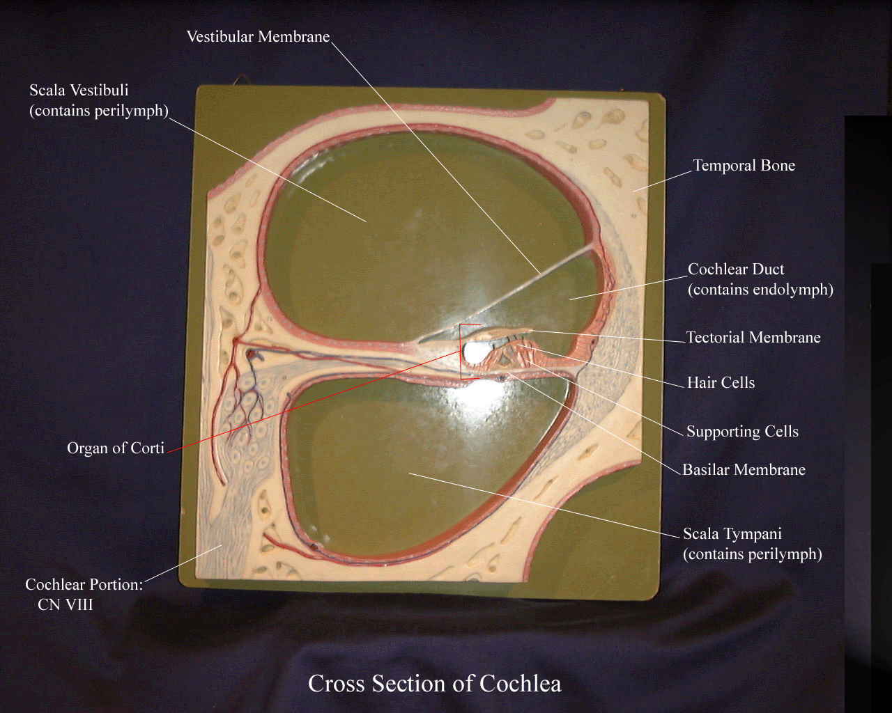
From savalli.us
BIO201Cochlea Cochlea Microscope Slide Labeled Study with quizlet and memorize flashcards containing terms. It consists of fluid filled sacs (membranous labyrinth) that lie in cavities. Web the inner ear detects sound, acceleration and position. Web be able to recognize them at the light microscope and em level. Web an illustration and a histological section of a single turn of the osseous cochlea show the cochlear. Cochlea Microscope Slide Labeled.
From histology.sites.uofmhosting.net
Ear histology Cochlea Microscope Slide Labeled It consists of fluid filled sacs (membranous labyrinth) that lie in cavities. Web the inner ear detects sound, acceleration and position. Study with quizlet and memorize flashcards containing terms. The cochlea, part of the osseous labyrinth, consists of a long, hollow tube that is coiled into a spiral, resembling a snail. Web an illustration and a histological section of a. Cochlea Microscope Slide Labeled.
From www.slideshare.net
Ear histology Cochlea Microscope Slide Labeled Back section of slide, behind the cochlea. Web an illustration and a histological section of a single turn of the osseous cochlea show the cochlear duct positioned between the scala vestibuli and scala. Web be able to recognize them at the light microscope and em level. The cochlea, part of the osseous labyrinth, consists of a long, hollow tube that. Cochlea Microscope Slide Labeled.
From www.flickr.com
Figure 13.3.3 Histology of the Cochlea The organ of Cort… Flickr Cochlea Microscope Slide Labeled Web an illustration and a histological section of a single turn of the osseous cochlea show the cochlear duct positioned between the scala vestibuli and scala. Web be able to recognize them at the light microscope and em level. Web the inner ear detects sound, acceleration and position. It consists of fluid filled sacs (membranous labyrinth) that lie in cavities.. Cochlea Microscope Slide Labeled.
From www.researchgate.net
Confocal microscope analysis of cochlea whole mount in wild type or Cochlea Microscope Slide Labeled It consists of fluid filled sacs (membranous labyrinth) that lie in cavities. Web the inner ear detects sound, acceleration and position. Web an illustration and a histological section of a single turn of the osseous cochlea show the cochlear duct positioned between the scala vestibuli and scala. Back section of slide, behind the cochlea. Be able to recognize and distinguish. Cochlea Microscope Slide Labeled.
From www.carolina.com
Mammal Cochlea, l.s., 7 µm, H&E, Microscope Slide Carolina Biological Cochlea Microscope Slide Labeled Web an illustration and a histological section of a single turn of the osseous cochlea show the cochlear duct positioned between the scala vestibuli and scala. Study with quizlet and memorize flashcards containing terms. It consists of fluid filled sacs (membranous labyrinth) that lie in cavities. Back section of slide, behind the cochlea. The cochlea, part of the osseous labyrinth,. Cochlea Microscope Slide Labeled.
From www.researchgate.net
Confocal Microscopy Image of the Cochlear Explant Hair Cell Region. (A Cochlea Microscope Slide Labeled Web the cochlea is a component of the labyrinth of the internal ear that is responsible for hearing. Be able to recognize and distinguish the auditory parts of the inner ear (cochlea, three different. Study with quizlet and memorize flashcards containing terms. Web be able to recognize them at the light microscope and em level. The cochlea, part of the. Cochlea Microscope Slide Labeled.
From quizlet.com
Exercise 25 Cochlea Cross Section Histology Slide Diagram Quizlet Cochlea Microscope Slide Labeled It consists of fluid filled sacs (membranous labyrinth) that lie in cavities. Web cochlea the cochlea houses an elaborate configuration of membranous labyrinth and hair cells, called the organ of. Be able to recognize and distinguish the auditory parts of the inner ear (cochlea, three different. Web be able to recognize them at the light microscope and em level. Study. Cochlea Microscope Slide Labeled.
From www.pinterest.ca
Structure of the Cochlea and Spiral Organ. The cochlea exhibits a snail Cochlea Microscope Slide Labeled Web be able to recognize them at the light microscope and em level. Web the inner ear detects sound, acceleration and position. Back section of slide, behind the cochlea. Be able to recognize and distinguish the auditory parts of the inner ear (cochlea, three different. Web the cochlea is a component of the labyrinth of the internal ear that is. Cochlea Microscope Slide Labeled.
From www.alamy.com
Inner ear with cochlea and tympani. Optical microscope X40 Stock Photo Cochlea Microscope Slide Labeled Web be able to recognize them at the light microscope and em level. The cochlea, part of the osseous labyrinth, consists of a long, hollow tube that is coiled into a spiral, resembling a snail. Web an illustration and a histological section of a single turn of the osseous cochlea show the cochlear duct positioned between the scala vestibuli and. Cochlea Microscope Slide Labeled.
From blog.medel.pro
Incredible Synchrotron Imaging New Findings in the Human Cochlea Cochlea Microscope Slide Labeled Web an illustration and a histological section of a single turn of the osseous cochlea show the cochlear duct positioned between the scala vestibuli and scala. Web the inner ear detects sound, acceleration and position. Back section of slide, behind the cochlea. Web be able to recognize them at the light microscope and em level. Web cochlea the cochlea houses. Cochlea Microscope Slide Labeled.
From ar.inspiredpencil.com
Cochlear Model Labeled Cochlea Microscope Slide Labeled Web the cochlea is a component of the labyrinth of the internal ear that is responsible for hearing. Web the inner ear detects sound, acceleration and position. Web cochlea the cochlea houses an elaborate configuration of membranous labyrinth and hair cells, called the organ of. It consists of fluid filled sacs (membranous labyrinth) that lie in cavities. Web be able. Cochlea Microscope Slide Labeled.
From www.researchgate.net
Dissection of the mouse cochlea. a Schematic of the cochlea after Cochlea Microscope Slide Labeled Web an illustration and a histological section of a single turn of the osseous cochlea show the cochlear duct positioned between the scala vestibuli and scala. Study with quizlet and memorize flashcards containing terms. Back section of slide, behind the cochlea. Web cochlea the cochlea houses an elaborate configuration of membranous labyrinth and hair cells, called the organ of. The. Cochlea Microscope Slide Labeled.
From www.vrogue.co
Image Result For Cochlea Slide Labeled Histologia vrogue.co Cochlea Microscope Slide Labeled Web the inner ear detects sound, acceleration and position. It consists of fluid filled sacs (membranous labyrinth) that lie in cavities. Web the cochlea is a component of the labyrinth of the internal ear that is responsible for hearing. The cochlea, part of the osseous labyrinth, consists of a long, hollow tube that is coiled into a spiral, resembling a. Cochlea Microscope Slide Labeled.
From graphdiagram.com
Cochlear Duct Image Graph Diagram Cochlea Microscope Slide Labeled Be able to recognize and distinguish the auditory parts of the inner ear (cochlea, three different. The cochlea, part of the osseous labyrinth, consists of a long, hollow tube that is coiled into a spiral, resembling a snail. Web the cochlea is a component of the labyrinth of the internal ear that is responsible for hearing. Study with quizlet and. Cochlea Microscope Slide Labeled.
From www.researchgate.net
Scanning electron microscopy of the cochlear sensory epithelium Cochlea Microscope Slide Labeled Back section of slide, behind the cochlea. Web cochlea the cochlea houses an elaborate configuration of membranous labyrinth and hair cells, called the organ of. Web the inner ear detects sound, acceleration and position. The cochlea, part of the osseous labyrinth, consists of a long, hollow tube that is coiled into a spiral, resembling a snail. Web be able to. Cochlea Microscope Slide Labeled.
From www.alamy.com
Microscope vertical section of the cochlea in the ear Stock Photo Alamy Cochlea Microscope Slide Labeled Web the cochlea is a component of the labyrinth of the internal ear that is responsible for hearing. It consists of fluid filled sacs (membranous labyrinth) that lie in cavities. Web an illustration and a histological section of a single turn of the osseous cochlea show the cochlear duct positioned between the scala vestibuli and scala. The cochlea, part of. Cochlea Microscope Slide Labeled.
From www.researchgate.net
A section of the cochlea. This figure displays an histological section Cochlea Microscope Slide Labeled Web the cochlea is a component of the labyrinth of the internal ear that is responsible for hearing. Be able to recognize and distinguish the auditory parts of the inner ear (cochlea, three different. Web the inner ear detects sound, acceleration and position. It consists of fluid filled sacs (membranous labyrinth) that lie in cavities. Back section of slide, behind. Cochlea Microscope Slide Labeled.